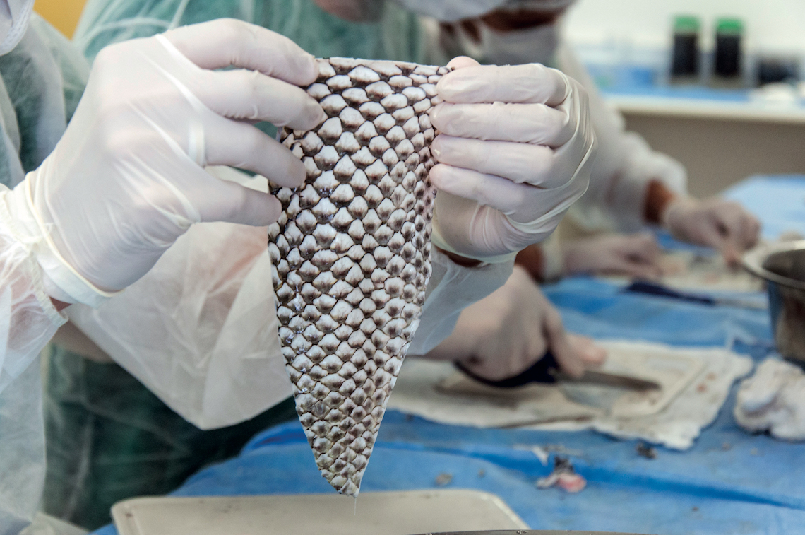
Tilapia fish skin has shown promise as a stable and useful biological dressing that can be used to treat wounds and burns. However, the appropriate sterilization technique of the Tilapia fish skin is crucial before its clinical application. The normal way of sterilizing things has to get rid of harmful germs while leaving behind the biochemical and structural features that might make the dressing less effective. This study looked into and compared how well three different disinfectants worked on the skin of tilapia fish. The three agents tested were chlorhexidine gluconate (CHG), povidone iodine (PVP-I), and silver nanoparticles (25% CE%BCg/mL) (AgNPs). The tests were done three times, at 5, 2010, and 2015 minutes, and were based on the number of microbes present, the histological findings, and the properties of the skin. However, among the methods used for sterilization, AgNPs demonstrated rapid and complete antimicrobial activity, with a 90% decrease in the growth of microbes on the fish skin during the treated times. Furthermore, AgNPs did not impair the cellular structure or collagen fibers content of the fish skin. However, CHG and PVP-I caused alterations in the collagen content. This study showed that treating Tilapia fish skin with AgNPs made the skin sterile while keeping the histological properties and structural integrity. It was found that this method of sterilizing Tilapia fish skin works well and quickly. It could be used as a biological dressing.
Biological dressings, like amnion, allografts, xenografts, bioengineered tissues, and cultured wound healing cells, are now being used to treat burns and skin wounds directly on the skin (1). Collagen dressings are widely used because they have many useful properties, such as low antigenicity, increased inflammation and hemostasis, and faster fibroplasia and epithelization (2). Because of the risk of disease transmission or religious and cultural concerns, collagen dressings made from cow and pig skin or chicken waste should not be used (3, 4). The skin of marine fish is a good source of collagen. It also has antibacterial and antioxidant properties thanks to omega-3 polyunsaturated fatty acids, especially eicosapentaenoic acid (EPA) and docosahexaenoic acid (DHA). Thus, it is unlikely to be a source of infectious disease (3, 5–8).
It was thought that Nile Tilapia (Oreochromis niloticus) skin could be used to treat burns and wounds because it is similar to human skin in terms of collagen, histology, and mechanics (9–11). Tilapia skin is a readily available, quality, safe, inexpensive material that is easy to apply (12). Nile tilapia skin is also very biocompatible because the collagen extract is a type I biocompatible collagen that could be used as a biomedical material in clinical regenerative medicine (13–16).
To make sure the patient is healthy and the wound heals properly, wound dressings must be sterilized before they are used on a patient. Ordinary methods used to sterilize skin grafts are ethylene oxide gas sterilization, irradiation, and chemical sterilization (17–19). Ethylene oxide is also a toxic gas that can cause harmful reactions that could be dangerous for the patient (20, 21). Irradiation sterilization of skin grafts made the structure of the skin matrix worse because it can break the peptide bonds within the collagen molecules or cross-link the collagen matrix (22). Mammal skin grafts need to be sterilized with harsh chemicals, but tilapia skin grafts need to be processed gently because the colony forming units found in the skin samples showed that there was a normal, non-infectious microbiota (23) However, microorganisms like Pseudomonas aeruginosa, Aeromonas sobria, Klebsiella pneumonie, Enterococus faecalis, Aeromonas hydrophila, Proteus mirabilis, Globicatella sanguinis, Aeromonas veronii, Streptococcus uberis, Candida parapsilosis, and Streptococcus suis may be on the fish skin. These microorganisms may have come from the water or other sources (23, 24).
People often use chlorhexidine and povidone iodine to clean and sterilize grafting procedures because they kill Gram-negative, Gram-positive, and fungi bacteria and fungi by disrupting the cell membrane (17, 25–28). Nanoparticles of silver have been used for a long time to kill microbes. They are used to make silver-based wound dressings and to clean wound grafts (19, 29).
A good way to sterilize must get rid of or greatly reduce the number of microbes that are present without changing the skin grafts’ structure, bioactive makeup, or biocompatibility (17–19). As far as the author knows, there aren’t many studies that look at how to sterilize fish skin and what effects that has on structure or function (17). The study aims to compare the number of microbes, the properties of collagen, and the histology of fish skin that has been sterilized three times and in three different ways.
The skin of a fresh Nile Tilapia (Oreochromis niloticus) fish (700 ± 40 gm, 22 ± 3 cm standard length) was used in this study. The fish came from the Aquatic Medicine Units tanks at Assiut University’s Faculty of Veterinary Medicine in Assiut, Egypt. According to the guidelines for euthanasia from the American Association of Zoo Veterinarians (AAZV), the fish were killed by cutting off their heads. Sharp and blunt dissection was used to get fresh skin samples from the muscles underneath. The samples were then washed with a sterile normal saline solution to get rid of any blood or other impurities. After putting the skin samples in a clean normal saline solution, they were cut into 3 cm by 3 cm strips. Right away, three different sterilization methods were used on tilapia skin strips, with three skin samples used in triplicate for each treatment. Each method was applied three times, at 5, 10, and 15 minute intervals, as shown below.
Three types of sterilizing agents were used: chlorhexidine gluconate 4% (CHG) from Hibiclens M.C. Health Care in Norcross, GA; povidone iodine 2010 (PVP-I) from Betadine, El-Nile Co.; and for Pharmaceutical and Chemical Industries, Egypt), and silver nanoparticles (AgNPs) (25 μg/mL). The skin strips from tilapia were randomly split into four groups, with three skin strips in each. These groups were CHG, PVP-I, and AgNPs. Each group was then incubated for different contact time points (5, 10, and 15 min). In addition to a control group (C) treated with normal saline for the three different times. Following each sterilization treatment at the times shown, the tilapia skin strips were examined for bacteria and tissue afterward.
As someone who loves experimenting with unique ingredients in the kitchen, I’m always interested in sourcing high-quality sterilized tilapia skin This fish skin has become prized for its versatility – it can be used for cooking, skin care, arts and crafts, and more However, finding reliably sterilized tilapia skin is not always straightforward. Through trial and error, I’ve discovered the best places to purchase high quality sterilized tilapia skin.
What Exactly is Sterilized Tilapia Skin?
Tilapia skin goes through a sterilization process to eliminate any bacteria and pathogens, rendering it safe for external use. The skin is harvested from the abundant Nile tilapia fish, then thoroughly washed, sterilized and packed.
Proper sterilization involves treatment with gamma irradiation or electron beam radiation. This kills microorganisms without compromising the skin’s collagen structure. The end result is tilapia skin that is reusable, durable and sterile.
Sterilized tilapia skin should have a translucent, pearly white appearance and feel smooth yet strong. It is sold dried, refrigerated or frozen. High standards of sterility, purity and freshness are essential when sourcing tilapia skin.
Where to Purchase Sterile Tilapia Skin
Here are my top recommended sources for buying quality sterilized tilapia skin
1. Online Retailers
A number of online retailers offer sterilized tilapia skin for reasonable prices. Two reputable options are:
-
RawSuperFoods.com – Good source for frozen tilapia skin. Ships in insulated coolers with dry ice.
-
Tilapia Depot – Sells dried tilapia skin. Offers bulk discounts. Fast shipping.
I recommend carefully vetting any online tilapia skin source. Check for proper sterilization methods and freshness guarantees before purchasing.
2. Asian Groceries and Fish Markets
Asian food stores and fish markets sometimes carry sterilized tilapia skin in the frozen section. Brands like Golden Prize are a good bet.
Inspect the packaging closely for sterilization details before buying. Freshness is also key – the skin should not have any off-odors or visible decay.
3. Restaurant Suppliers
Many restaurant suppliers offer sterilized tilapia skin for sushi restaurants and other commercial kitchens. Check suppliers like Cheney Brothers or Jade Asian Foods.
Call ahead to check current availability and sterilization methods. Buying in bulk yields the best price per pound but requires large minimum orders.
4. Skin Care Facilities
Some skin care clinics, salons and plastic surgeon offices purchase sterilized tilapia skin in bulk quantities for procedures.
Ask nicely and they may be willing to sell smaller packages of unused tilapia skin. This ensures proper sterilization and freshness.
5. University Labs and Research Centers
University biology, zoology and marine science departments occasionally use sterilized tilapia skin for research.
Getting in touch with their labs to ask about buying excess unused tilapia skin is worth a shot. The sterilization will likely be up to scientific standards.
6. Tilapia Farmers
If you happen to live near a tilapia farm, check if they sterilize skin for sale. This yields the freshest option, as you cut out supply chain lag.
Be sure to confirm their sterilization process meets food safety standards before using the skin.

Preparation of Silver Nanoparticles
Stable silver nanoparticles (AgNPs) <100 nm were synthesized in a typical one-step protocol as described before (31). Briefly, 1. 0 g of soluble starch was added to 100 mL of deionized water and heated until complete dissolution. One milliliter of 100 mM aqueous solution of silver nitrate (AgNO3) crystal (ACS AgNO3, F. W 169, 87 Gamma laboratory chemicals, Sigma-Aldrich, Germany, Assay: Min 99. 0%) was added and stirred well. This mixture was put into a dark glass bottle and autoclaved. The resulting solution was clear yellow in color indicating the silver nanoparticles formation.
The AgNPs stock solution was kept in dark glass bottles at room temperature (25°C) and out of direct sunlight. The concentration and size of the particles were measured before using. We used a transmission electron microscope (TEM) (JEOL-JEM-100CX II) at the Electron Microscopy Unit, Assiut University to look at the shape and size of AgNPs (Supplementary Figure 1). The Graphite Furnace Atomic Absorption Model 210VGP at the Faculty of Science, Assiut University was used to measure the amount of AgNPs stock.
Microbiological evaluation of the fish skin strips was performed according to APHA (32) and Qazi et al. (33). An area of 3 × 3 cm2 was aseptically swabbed for microbial count. We used Plate count agar (Ref. -M091A), Manitol Salt Agar (Oxoid CM0085), MacConkey Agar OMRI W/O Crystal violet (Biolife) and DifcoTM Potato Dextrose Agar (Ref. 213400) to count the total number of viable bacteria, Staphylococci, Coliforms, yeasts, and molds. The Pour plate method was used for the enumeration of colony-forming units (CFU/cm2) on Plate Count Agar. The swab was transferred immediately into 10 ml sterile nutrient broth. One milliliter from the broth was transferred aseptically into a sterilized petri-dish in triplicate. A water bath was used to cool ten to fifteen milliliters of the dissolved Plate count agar media to 44–46°C, and this was then poured into each plate in a clean way. A circle was drawn around the dish several times to make sure that the sample and medium were all the same. The plate was left until the medium hardened, and then it was put upside down in an incubator set to 37°C for 24 hours. After the incubation period, the number of each replicate sample was counted, and the mean was found by taking that number and multiplying it by the opposite of the dilution. The resulting colonies expressed as CFU/ml. The same technique was used for Staphylococci, Coliforms, Yeasts and Molds count. The number of Staphylococcal bacteria was grown on Manitol Salt Agar “Oxoid CM0085” at 37°C for 24 hours. The number of Coliform bacteria was grown on MacConkey Agar OMRI W/O Crystal Violet “Biolife at 37°C for 18–24 hours. The number of Yeasts and Molds was grown on DifcoTM Potato Dextrose Agar– Ref 213400 and incubated at 28 ± 2°C for 24–72 hours.
To figure out how well each sterilization method worked, we compared the number of bacteria before and after each treatment for each contact time using the following equation:
The effectiveness of disinfection is 20% (C0%20%E2%80%93%20C)/C0%20%C3%97%20100, where (C0) is the initial bacterial count (control negative) and (C) is the count of bacteria after a certain amount of time of contact with the sterilizing agent (34)
Specimens (0. 5 × 0. Skin strips (5 cm) of tilapia were taken from each group after each sterilization treatment at different times and fixed in 10% neutral buffered formalin. The formalin-fixed skin samples were routinely processed and embedded in paraffin. Then, 5 μm thick slices were made and colored with Mayers hematoxylin (Merck, Darmstadt, Germany) and eosin (Sigma, Missouri, USA). After that, the slides were looked at under a microscope, and the histological evaluation was done on coded samples without anyone knowing who did it. Sections from the control-treated group were then compared. Histological evaluation to assess integrity and organization of collagen fibers was based on 0–3 scale (35, 36). The collagen integrity scores are 0 for long fibers that don’t break, 1 for slightly broken fibers, 2 for moderately broken fibers, and 3 for severely broken fibers. Collagen organization scores: 0 means the collagen is tight and straight, 1 means it’s a little loose and waves, 2 means it’s moderately loose, waves, and crosses over itself, and 3 means there is no clear pattern.
Negative Image Study Using CMEIAS Color Segmentation
Better computer technology has been used to process colors by separating pixels from background objects from pixels from foreground objects (38, 39). This method was especially useful for this analysis because the colors were so complex.
Fishy But True: Tilapia Skin Heals Burns. Plastic Surgery Hot Topics with Rod J. Rohrich, MD
FAQ
How is tilapia skin sterilized?
What are the disadvantages of tilapia skin for burns?
What is tilapia skin made of?
Do fish skin bandages help heal wounds?
